Clinical Image
Intra-Operative View of Arterial Branch from the Aorta to the Left Lower Lobe during VATS Lobectomy for Intra- Pulmonary Sequestration
ElKhayat H1*, Sallam M1, Khalil M2 and Elminshawy A2
1Department of Cardiothoracic Surgery, Assiut University Heart Hospital, Egypt
2Department of Cardiothoracic Surgery, Aberdeen Royal Infirmary, United Kingdom
*Corresponding author: Hussein ElKhayat, Department of Cardiothoracic Surgery, Assiut University Heart Hospital, Egypt
Published: 01 Dec, 2017
Cite this article as: ElKhayat H, Sallam M, Khalil M,
Elminshawy A. Intra-Operative View
of Arterial Branch from the Aorta to
the Left Lower Lobe during VATS
Lobectomy for Intra-Pulmonary
Sequestration. Clin Surg. 2017; 2: 1790.
Clinical Image
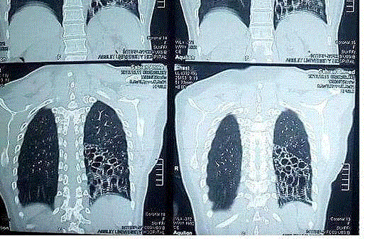
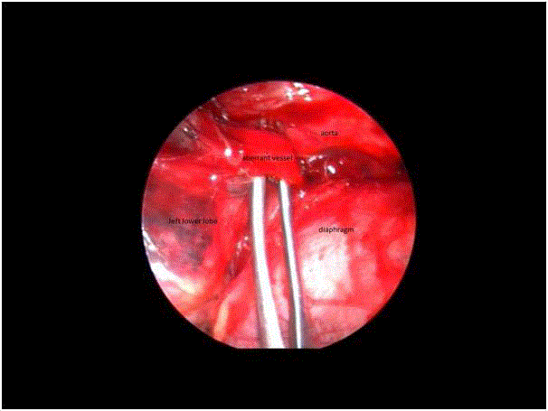
A 50-year old female presented to our unit with recurrent chest infections since childhood. CT showed extensive bronchiectasis of the left lower lobe, and decision was made to remove the bonchiectatic lobe through VATS lower lobectomy. An incidental finding of an arterial branch supplying the lobe from the thoracic aorta during dissection of the inferior pulmonary ligament confirmed this to be a case of intrapulmonary sequestration (Figure 1). The aberrant branch was divided and the lobe removed in the usual manner. This case reiterates the importance of care while dividing the inferior pulmonary ligament during VATS removal of lower lobes with recurrent infection, as inadvertent injury or transaction of such aberrant vessels could result in fatal haemorrhage (Figure 2). Aberrant vessels to sequestrated lobes arise directly from the aorta, and if completely transacted, the proximal end could retract below the diaphragm, making access and control very difficult even with the chest opened.


